Description
Mammal Pseudostratified Ciliated Columnar Epithelium Slide, 7 µm, H&E
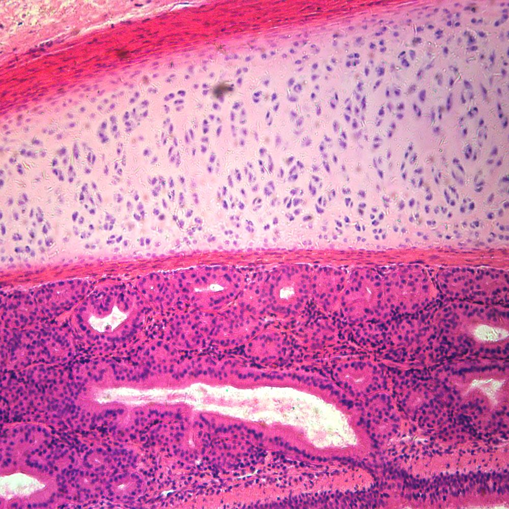
Taken from a section of trachea and stained with hematoxylin and eosin, this pseudostratified ciliated columnar epithelium slide is great for the study of epithelial tissues. As its name suggests, the tissue appears to consist of multiple cell layers due to cell nuclei appearing at different levels. However, the tissue actually consists of 1 layer of columnar cells (pseudo-stratified). Students can also observe the small cilia projecting from the cells, aiding in transporting and trapping substances in the trachea and respiratory tract.
The tissue is classified by the number of cell layers it has (simple=1 cell layer, stratified=more than 1 cell layer) and the shape of the cells (squamous=flat, cuboidal=cube-shaped, columnar=column-shaped).