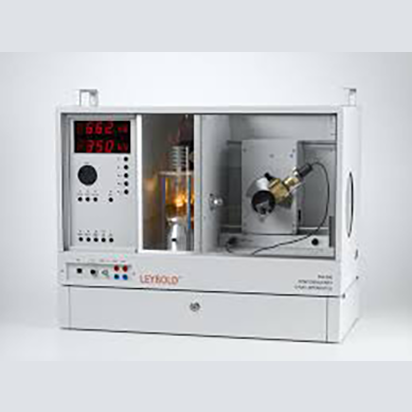
LD Didactic X-Ray Apparatus


Category: Tag:

Description

Setting New Standards in Resolution & Intensity
LEYBOLD’s X-RAY Apparatus impresses with its superior safety concept, and its continuously improved capabilities and features. Six different X-ray tubes (with different anode materials) are available, which is a unique feature for an educational X-ray apparatus.

This X-RAY apparatus is setting new standards in resolution and intensity in the field of education worldwide. Not only the high resolution of the Bragg spectra is impressive, but also the new high-resolution X-ray image sensor, the reliable X-ray energy detector and the gold tube.

The modular structure of the system enables both a low-cost introduction (FUNDAMENTAL Experiments) and advanced applications (PROFESSIONAL Experiments) for several different test subjects.

“We have implemented the LEYBOLD cabinet X-ray system in both undergraduate and graduate labs and we are very happy with the results. The students really enjoy being able to see all components of the X-ray tube. AYVA set us up quickly and we haven’t had any issues since. The modular cabinet X-ray system is customizable for a variety of experiments from materials analysis, to dosimetry, to even imaging. We are looking forward to working with AYVA to expand the system capabilities in the future.”
James Gräfe
Physics Professor
Ryerson University

FUNDAMENTALS

  • Radiography
  • X-ray photography
  • Ionisation and Dosimetry
  • Attenuation of X-ray beams

CRYSTALLOGRAPHY

  • Bragg: Determining the lattice constants of monocrystals
  • Laue: Investigating the lattice structure of monocrystals
  • Debye-Scherrer: Determining the lattice planespacings of polycrystalline powder samples

APPLICATIONS

  • Radiology
  • Mineralogy
  • Radiation protection
  • X-ray fluorescence analysis
  • Non-destructive material analysis
  • Non-destructive testing
  • Computed tomography also in 3D

ATOMIC PHYSICS

  • Bragg: Diffraction on X-ray beams on a monocrystal
  • Investigating the energy spectrum of an X-ray tube
  • Duane-Hunt: Determination of h from the threshold wavelength
  • Energy-dependent absorption, K- and L-edges
  • Moseley’s law and determination of the Rydberg constants
  • Fine structure of X-ray spectra
  • X-ray fluorescence
  • Compton effect on X-ray radiation


PDF (Instruction Sheet) PDF (Instruction Sheet) [554 800] X-ray apparatus
PDF (Experiment description) PDF (Experiment description) P6.3.1.1 Fluorescence of a luminescent screen due to X-rays
PDF (Experiment description) PDF (Experiment description) P6.3.1.2 X-ray photography: Exposure of film stock due to X-rays
PDF (Experiment description) PDF (Experiment description) P6.3.1.3 Detecting X-rays using an ionization chamber
PDF (Experiment description) PDF (Experiment description) P6.3.1.4 Determining the ion dose rate of the X-ray tube with molydenum anode
PDF (Experiment description) PDF (Experiment description) P6.3.1.5 Investigation of an implant model (de)
PDF (Experiment description) PDF (Experiment description) P6.3.1.6 Influence of a contrast medium on the absorption of X-rays
PDF (Experiment description) PDF (Experiment description) P6.3.2.1 Investigating the attenuation of X-rays as a function of the absorber material and absorber thickness
PDF (Experiment description) PDF (Experiment description) P6.3.2.2 Investigating the wavelength dependency of the attenuation coefficient
PDF (Experiment description) PDF (Experiment description) P6.3.2.3 Investigating the relationship between the attenuation coefficient and the atomic number Z
PDF (Experiment description) PDF (Experiment description) P6.3.5.1 Recording and calibrating an X-ray energy spectrum
PDF (Experiment description) PDF (Experiment description) P6.3.5.2 Recording the energy spectrum of a molybdenum anode
PDF (Experiment description) PDF (Experiment description) P6.3.5.3 Recording the energy spectrum of a copper anode
PDF (Experiment description) PDF (Experiment description) P6.3.5.4 Investigation of the characteristic spectra as a function of the element’s atomic number: K-lines
PDF (Experiment description) PDF (Experiment description) P6.3.5.5 Investigation of the characteristic spectra as a function of the element’s atomic number: L-lines
PDF (Experiment description) PDF (Experiment description) P6.3.5.6 Energy-resolved Bragg reflection in different orders of diffraction
PDF (Experiment description) PDF (Experiment description) P6.3.6.1 Fine structure of the characteristic X-ray radiation of a molybdenum anode
PDF (Experiment description) PDF (Experiment description) P6.3.6.11 High-resolution fine structure of the characteristic X-ray radiation of a molybdenum anode (de)
PDF (Experiment description) PDF (Experiment description) P6.3.6.12 High-resolution fine structure of the characteristic X-ray radiation of a copper anode (de)
PDF (Experiment description) PDF (Experiment description) P6.3.6.13 High-resolution fine structure of the characteristic X-ray radiation of an iron anode (de)
PDF (Experiment description) PDF (Experiment description) P6.3.6.14 High-resolution fine structure of the characteristic X-ray radiation of a silver anode (de)
PDF (Experiment description) PDF (Experiment description) P6.3.6.15 High-resolution fine structure of the characteristic X-ray radiation of a tungsten anode (de)
PDF (Experiment description) PDF (Experiment description) P6.3.6.17 High-resolution fine structure of the characteristic X-ray radiation of a gold anode (de)
PDF (Experiment description) PDF (Experiment description) P6.3.6.2 Fine structure of the characteristic X-ray radiation of a copper anode
PDF (Experiment description) PDF (Experiment description) P6.3.6.3 Fine structure of the characteristic X-ray radiation of an iron anode
PDF (Experiment description) PDF (Experiment description) P6.3.6.4 Fine structure of the characteristic X-ray radiation of a silver anode (de)
PDF (Experiment description) PDF (Experiment description) P6.3.6.5 Fine structure of the characteristic X-ray radiation of a tungsten anode
PDF (Experiment description) PDF (Experiment description) P6.3.6.7 Fine structure of the characteristic X-ray radiation of a gold anode (de)
PDF (Experiment description) PDF (Experiment description) P6.3.7.1 Compton effect: verifying the energy loss of the scattered X-ray quantum
PDF (Experiment description) PDF (Experiment description) P6.3.7.2 Compton effect: Measurement the energy of the scattered photons as a function of the scattering angle
PDF (Experiment description) PDF (Experiment description) P6.3.8.1 Measurement and presentation of a computed tomogram with the computed tomography module
PDF (Experiment description) PDF (Experiment description) P7.1.2.1 Bragg reflection: determining the lattice constants of monocrystals
PDF (Experiment description) PDF (Experiment description) P7.1.2.2 Laue diagrams: investigating the lattice structure of monocrystals
PDF (Experiment description) PDF (Experiment description) P7.1.2.3 Debye-Scherrer photography: determining the lattice plane spacings of polycrystalline powder samples
PDF (Experiment description) PDF (Experiment description) P7.1.2.4 Debye-Scherrer Scan: determining the lattice plane spacings of polycrystalline powder samples
PDF (Experiment description) PDF (Experiment description) P7.5.1.1 Application of X-ray fluorescence for the non-destructive analysis of the chemical composition
PDF (Experiment description) PDF (Experiment description) P7.5.1.2 Determination of the chemical composition of a brass sample by X-ray fluorescence analysis
PDF (Experiment description) PDF (Experiment description) C3.6.5.1 X-ray fluorescence analysis of chemical composition
PDF (Experiment description) PDF (Experiment description) C3.6.5.2 The chemical composition of a brass specimen